Neurobiology of addiction, UTC for healthcare professionals
| To read the full text of the in-depth manual of the UTC for healthcare professionals, please visit: https://www.issup.net/node/7376 |
Please scroll down to read the full text and watch the video at the bottom.
Neurons
Neurons are the functional units of the nervous system, considered as “the cells of the mind.”They specialize in detecting changes in the environment and in responding to them; they also coordinate the functions of the organism itself. In broad terms, they consist of a cell body or soma and extensions (projections) of two types: dendrites and axons. To understand the importance of these structures, note that the body nerves are large groups of many axons. Neurons communicate through their extensions. The areas where this communication takes place are the synapses.
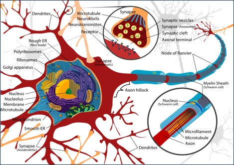
Synapses
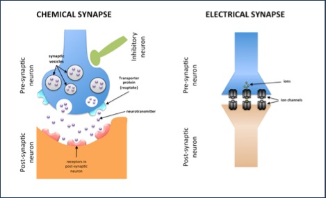
A single neuron can be connected to many others by multiple synapses. As an example, we shall describe a simplified model of communication between two neurons. The most common synapses are chemical, although there are also electrical synapses. Both types have three common elements: the presynaptic neuron, the synaptic cleft or space, and the postsynaptic neuron. These names are used to refer to the flow of information traveling between cells. In this example, the presynaptic neuron transmits the message to the post-synaptic neuron.
In chemical synapses, at the end of the presynaptic axon, there are vesicles or reservoirs with the molecules that will take the signals to the receptors on the postsynaptic neuron membrane.
Like all cells, neurons have a cellular membrane that seals them off from the environment. Thanks to this separation, inside the neuron there is a higher concentration of potassium ions and proteins, while on the outside there are high concentrations of sodium and chlorine.
To produce their effects, chemical messengers bind to cellular receptors. This binding property is known as affinity, while efficacy refers to their capacity to generate physiological effects. Agonist substances have high affinity and high efficiency, while antagonist substances have high affinity and low efficiency.
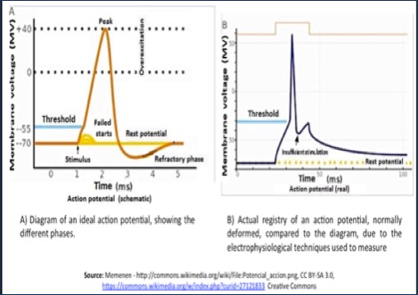
Action potential
Neurons understand and use an electrical language based on the charges from the ions and molecules that are inside and outside the cellular membrane. When a neuron is “quiet or silent,” its internal environment has more negative electric charges than the outside. These conditions change abruptly when the neuron interacts with others. A single neuron can communicate with many others at the same time and can "understand" the final message because it integrates the multiple electrical signals it receives. Based on the changes of permeability to specific ions of its cellular membrane, neurons decode differences in the distribution of internal electrical charges. A positively charged environment produces a wave of electrical information called action potential, which propagates rapidly and in all directions within the cell and also through the axon. If this action potential reaches an electrical synapse, the current passes directly to the postsynaptic neuron; but if it is a chemical synapsis, the change in the electrical charge opens some pores in the membrane of the neuron through which the calcium ion enters. This higher concentration of calcium inside the presynaptic neuron sets off the start of the neurotransmission.
Neurotransmission
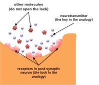
Communication between neurons is highly efficient, either among them or with other types of cells through chemical or physical mechanisms, but rarely by direct physical connections (called gap junctions). In general, the most common way of doing this is via indirect chemical contacts (chemical synapses), which is where neurotransmission occurs. During this process, the action potentials enable signals and messages to spread among the neurons and the neuronal networks. The neurotransmitters released into the synaptic cleft interact with specific receptors, which causes changes in the postsynaptic neuron. Due to their affinity and specificity, the binding of the neurotransmitter molecules with their receptors is comparable to the way a door key operates in the lock. The neuronal receptors recognize the tridimensional configuration and chemical characteristics of neurotransmitter molecules. Neurons release the neurotransmitter into the synaptic space via the process of exocytosis, which consists of the following steps: 1) docking, 2) priming, 3) vesicular fusion/exocytosis, 4) endocytosis mediated by receptosomes and clathrins, 5) intracellular translocation, 6) endosomal fusion, 7) gemmation with the budding of new vesicles, 8) active storage of the neurotransmitter, and 9) cytoplasmic translocation to begin the cycle again. The molecular compound formed with the union of the neurotransmitter and its receptor remains active for a short period. There are two known ways in which the action of the neurotransmitter can be halted: reuptake and degradation. In reuptake, the presynaptic neuron uses membrane proteins (reuptake pumps or transporters) which collect the released neurotransmitter and reintroduces it into the vesicles of the presynaptic neuron, so that it can be used again in the future. In degradation, specific enzymes inactivate the neurotransmitter and convert it into a different compound. The presynaptic neurons send messages that, depending on the neurotransmitter released and the functions of the neurons involved, may stimulate or inhibit the postsynaptic neuron. The outcome is determined by the sum of the many stimuli that are received.
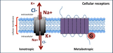
Receptors
Neurons have specialized protein structures that because of their specific interaction with other chemical substances serve the process of cellular communication. The substances that bind with receptors are known as “ligands.” For neuronal communication, we shall discuss two large groups of receptors:
Ionotropic– when activated by their ligand, the tridimensional structure and configuration of their proteins change, allowing the ions, whether negatively charged (chloride) or positively charged (calcium, potassium and sodium), to pass through. They are also known as ionic channels.
Metabotropic– are formed by long chains of amino acids that pass seven times through the cellular membrane. The interaction with their ligand activates or inhibits specific proteins (G proteins) linked to the cellular membrane, which are involved in the production of other molecules such as AMPc.
Neurotransmitters
Neurotransmitters are the interlocutors in conversations between neurons. In general terms, they are substances that have a simple chemical structure, grouped under three broad categories: amino acids, amines, and peptides. These substances intervene in the communication between neurons that control very different functions.
Dopamine– this catecholaminergic neurotransmitter participates in regulating movement, emotion, affect, and neuroendocrine communication. It is synthesized from the L-tyrosine amino acid. At least there are five different types of dopaminergic receptors; all coupled to G proteins and part of two pharmacological families: 1) D1-like family (subtypes D1 and D5) – stimulate the formation of AMPc, and 2) D2-like family (subtypes D2, D3, and D4) – inhibit the formation of AMPc, activate K+ channels and reduce the entry of Ca++ ions through voltage-dependent channels. There are different subtypes of dopaminergic receptors distributed throughout the Central Nervous System (CNS). In the Peripheral Nervous System, dopamine modulates the cardiac and renal functions, vascular tone and gastrointestinal motility. Dopamine and dopaminergic neuronal networks get much attention because of their critical role in illnesses and disorders such as Parkinson’s disease, schizophrenia, and drug use disorders. In the latter case, besides dopamine, there are other neurotransmitters involved.
GABA– the Gamma-Aminobutyric Acid is the primary inhibitory neurotransmitter in the CNS. Approximately 30-40% of neurons in the brain use it to communicate by means of the GABAA receptors (ionotropic) and GABAB (metabotropic). Five subunits form the GABAA receptors, which have at least eleven different structural sites for substances like barbiturates, benzodiazepines, ethanol, and others, whether agonists or competitive antagonists. Due to its structure, the conformation of the GABA molecule is flexible, which means that its various configurations may activate distinct GABA receptors.
Noradrenaline or norepinephrine– this catecholamine is a derivate product of the tyrosine amino acid. It takes part in alertness, level of consciousness, motivation, sensory perception, regulation of the sleep-wakefulness cycle, appetite, sexual behavior and neuromodulation of learning, memory and reward mechanisms; in most of these functions, it operates jointly with other neurotransmitters. There are two known classes of noradrenergic receptors: alpha and beta. The alpha1 are excitatory and exist in the brain, vascular smooth muscle, intestine, and cardiac muscle. The alpha2 have inhibitory effects and exist in the brain, vascular smooth muscle, gut, nerve endings, and platelets. There are three known types of beta receptors. The beta1 are excitatory and are present in the heart; beta2 receptors are present in smooth and striated muscle, the liver and lymphocytes, and also are excitatory. The beta3 are present in adipose tissue.
Serotonin– 5-hydroxytryptamine or 5-HT is an indolamine derived from the amino acid tryptophan. It is synthesized mainly in the raphe nuclei of the brainstem which projects throughout the central nervous system, with greater density in the basal ganglia and the limbic structures. Serotonin regulates a wide variety of effects mediated by binding to specific membrane receptors, which are present in both the Central and Peripheral Nervous Systems, and also in many other tissues of the intestine, cardiovascular system, and blood cells. There are up to seven members of the family of serotonin receptors (5-HT1 to 5-HT7) and several subtypes. The 5-HT receptor groups are coupled to G proteins, except for 5-HT3, which uses ion channels. The broad distribution of the many subtypes of 5-HT receptors in the CNS explains the wide variety of their actions. Serotonin takes part in regulating mood, excitation, impulsivity, aggression, appetite, and anxiety.
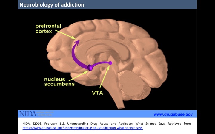
The Reward Pathway
Although different drugs have distinctive chemical properties, an element common to all is their capacity to produce positive reinforcing effects, which means that they cause an increase in the frequency of occurrence of the studied behavior (such as drug use), which tends to repeat. The first studies in this field come from the work of James Olds and Peter Milner. In 1954, these researchers conducted a study in rats, in which they observed that intracranial electrical stimulation of the hypothalamus and some associated regions might act as a reinforcer or reward for behaviors. Following their initial fortuitous observations, they used the paradigm of electrical self-stimulation in animal models to map the regions of the brain involved and discovered what they called the pleasure center. We know today that reinforcement comes from direct or indirect activation of the mesocorticolimbic dopamine system, which is formed by dopaminergic mesencephalic projections originating in the ventral tegmental area (VTA) and that project into the nucleus accumbens, the drugs’ principal target. The VTA neurons connect their axons to the nucleus accumbens, the striatum and the frontal cortex (structures that play a crucial role in motivation). The nucleus accumbens, a structural part of the limbic system, receives dopaminergic connections from the VTA, and glutamatergic connections from the lateral prefrontal cortex, the amygdala, and the hippocampus. The fact that the impulses coming from the cerebral cortex and the limbic system enter the nucleus accumbens shows the importance of this structure for the processes of motivation and reward produced by the repeated use of drugs.
Once we understand the formation of the neural pathways related to pleasure and reward, it is easier to comprehend how the artificial overstimulation that drugs produce in the nucleus accumbens causes the pleasurable effects that people who use drugs are seeking and makes them repeat this behavior. Drugs produce this due to different pharmacological properties and action mechanisms, discussed in the section about the effects of drugs. The brain is wired to ensure repetition of the activities that sustain life, by linking them with pleasurable, rewarding, or gratifying actions. Neural networks memorize which stimuli activate the reward system, fostering future repetition without actively thinking.
Some psychoactive substances can release two to ten times more dopamine in the reward circuit than the amount released by natural elements like eating and sexual intercourse. This consequence occurs almost immediately when smoking or injecting drugs, and their effects can last much longer than those produced by natural reinforcers. Such a big reward deeply motivates people to use drugs again and again.
There are three neuronal circuits mainly involved in the development of drug use disorders:
- Binge/intoxication: the reinforcing effects of the drugs can compromise reward neurotransmitters and associative mechanisms in the nucleus accumbens, and the stimulus-response habits that depend on the dorsal striatum. Two important neurotransmitters that mediate the rewarding effects of drugs are dopamine and opioid peptides.
- Withdrawal/negative affect: the negative emotional state of abstinence may compromise the activation of the extended amygdala. The main neurotransmitters involved in the negative reinforcement are the corticotropin-releasing factor, noradrenaline, and dynorphin. The extended amygdala projects mainly into the hypothalamus and brainstem.
- Preoccupation/anticipation (craving): Executive control is governed by the prefrontal cortex and includes the representation of contingencies, results, and their value and the subjective states (i.e., desire and, presumably, feelings) associated with drugs. The subjective effects called drug cravings in humans involve activation in studies of functional images of the orbital and anterior cingulate cortex and the temporal lobe, including the amygdala. Glutamate is an important neurotransmitter involved in the stage of craving and is located in the pathways of the frontal regions.
Drug use disorders are similar to other diseases, for example, cardiac conditions, that is, in both the regular and healthy functioning of underlying organs is interrupted, they have serious consequences, they are preventable, treatable and can last a lifetime without treatment.
People who use drugs experience alterations in brain functions such as a drastic decrease in the ability to perceive pleasure. Neuroimaging techniques allow demonstrating the evident differences of neuronal activity in the brains of drug users, in comparison with individuals who do not consume. As an example, methamphetamine users either undergo a significant reduction in dopamine transporters or a lower number of receptors that can receive signals. These neuroadaptive processes, named tolerance, lead to keep using drugs, again and again, trying to restore the normal dopamine functions, utilizing larger quantities of the substances to attain their psychoactive effects.
Related topics
- Classification of Drugs: https://www.issup.net/node/7521
- ISSUP members can join Networks to comment – Sign in or become a member